Packaging Details
Gross weight12 kg
Net weight7 kg
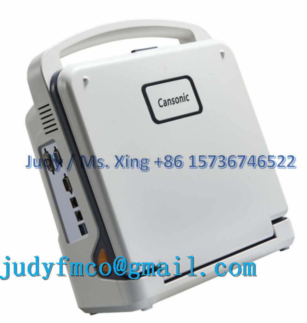
YJ-U60PLUS  3D Color Doppler
                --Full Digital Color Doppler Ultrasound Diagnostic System
  Â
Â
AppearanceSmart, compact  and clamshell design12 inch LED monitorBacklit operation panel, 8TGCFloating keyboardTwo active probe connectorsTwo probe holdersÂ
Probe
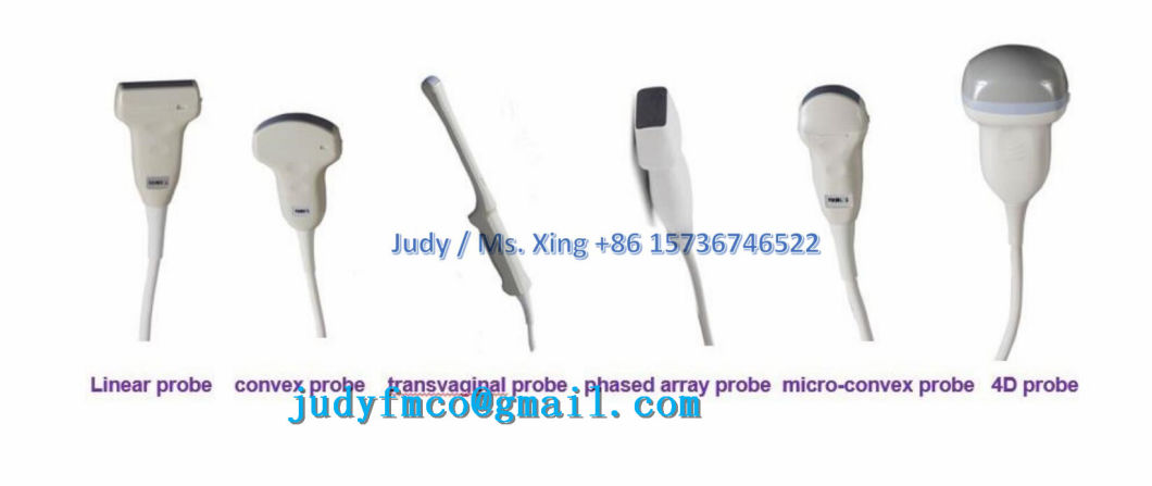
Transducer Types3.5Mhz convex probe (2.0/ 3.0/ 3.5/ 4.0/ 5.5Mhz)7.5Mhz linear probe (6.0/ 6.5/ 7.5/ 10.0/ 12.0Mhz)3.5Mhz micro convex probe (2.0/ 2.5/ 3.5/4.5/ 5.0Mhz)6.5Mhz transvaginal probe  (5.0/ 6.0/ 6.5/ 7.5/ 9.0Mhz)3.5Mhz phased array probe (2.1/ 3.0/ 3.5/ 4.0/ 5.0Mhz)4D volume probe (2.0/ 3.0/ 3.5/ 4.0/ 5.5Mhz)ApplicationsAbdomen, Obstetrics, Gynecology, Pediatrics,Small parts, Artery,  Superficial organ, Orthopedics, Cardiology, Musculoskeletal, Vascular, etcFunctionAuto Image Optimization Tissue Harmonic imagingiClear (Speckle Noise Reduction)iBeam (Spatial Compound Image)iZoom PIHI (Pulse-Inverse Harmonics Imaging)SA (Synthetic Aperture ultrasonic Imaging)Panoramic Image  (Option)Trapezoid Image (Option)Continuous Wave Doppler(Option)Â
Display modeB, B|B,4B, B|M,M,B|D,PW,B|PW, CFDuplex/Triplex modeCW (option)4D mode (option)Â
ZoomReal time zooming- 10 Steps:  ×1.0, ×2.0, ×3.0, ×4.0, ×5.0, ×6.0, ×7.0, ×8.0, ×9.0, ×10.0Selectable zooming positionÂ
FocusContinuous dynamic focus1~16 selectable transmit focusAcoustic lens focus1, 2, 3, 4 focusÂ
Memory
 Cineâ€memoryBâ€modeMâ€mode SSD (Solid State Disk) 64G
Â
Imaging Processing
B mode8â€step TGC slide pots- Gain: 0~100%
- Depth: 1.6~30cmFrequency: 5Â stepsDynamic range adjustable:Â 0~150dBEdge enhancement:0~7Persistence:0~7Chroma:0~6Grayscale:0~16- Power: 0~100%
- Noise reduction: 0-6iclear: off, 1, 2, 3, 4Â
MÂ mode
- Gain:Â 0~100%Sweep speed: 4Â steps- Maps:Â 0~16Chroma:0~6Â
CÂ mode
- Gain:Â 0~100%Pulse waveWall filter: 4Â stepsColor Maps:Â 0~7Package size: Â 8~15Color persistence:Â 0~7Threshold: 0-3Base line: 0-6Line density: Low and highSpatial filter: 0-3Â
PWÂ mode
- Gain:Â 0~100%Frequency: 5Â stepsPseudo color:0~6PRFd:1.0~6KHzBasic line: 7 stepsWall filter: 7Â steps- Spectrum mode: Refresh and Synchronize
 -   Sampling volume: 0.5-48mm
Â
Measurement &Â Calculation
Measurement
B mode (General) DistanceTrace LengthEllipse (area)Trace(area)AngleVolumeÂ
PW modeHR (heart rate)DistanceVelocityTimeÂ
Calculation
AbdomenLiverGallbladderPancreasSpleen UrologyKidneyUreterBladderAfter the urine bladderProstate GynecologyUterusCervixOvaryFollicle Â
Â
Early Obstetrics GSBPDCRLNT…
Later ObstetricsBPDHCACOFDFLTAD...
Small partsThyroidTestes
Â
MusculoskeletalHipÂ
Peripheral vascularIntimaArteryÂ
CardiologyDistanceAngleVolumeRVWd LVDd RVDd LVPWdRVWsLVDsRVDsLVPWsRV/LVAO…
Â
Physical Features
ConnectivityVideo out portDVI out portVGA out port2  USB portDICOM 3.0Â
Dimension
Â
|
 Full Digital Color Doppler Ultrasound Diagnostic System   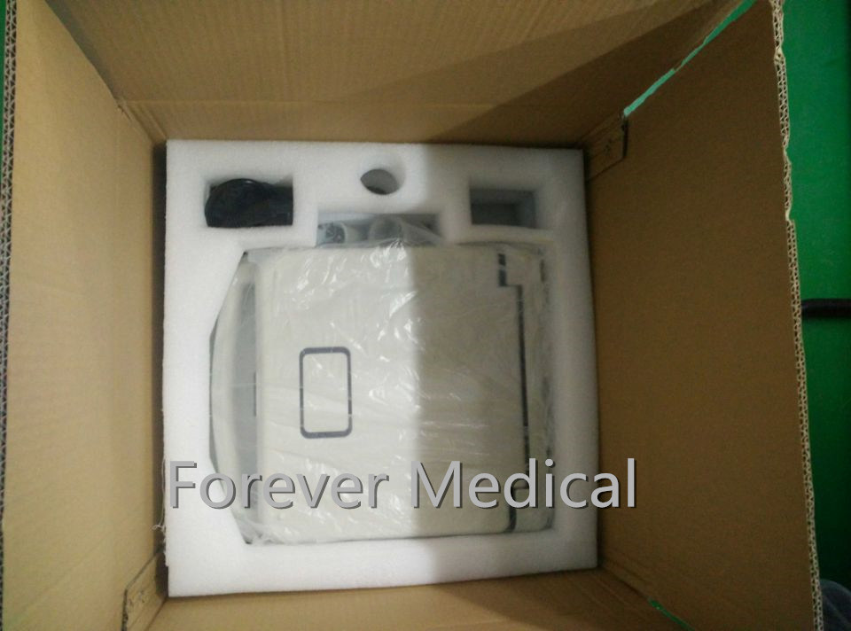   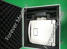 AppearanceSmart, compact  and clamshell design12 inch LED monitorBacklit operation panel, 8TGCFloating keyboardTwo active probe connectorsTwo probe holders. Probe Transducer Types3.5Mhz convex probe (2.0/ 3.0/ 3.5/ 4.0/ 5.5Mhz)7.5Mhz linear probe (6.0/ 6.5/ 7.5/ 10.0/ 12.0Mhz)3.5Mhz micro convex probe (2.0/ 2.5/ 3.5/4.5/ 5.0Mhz)6.5Mhz transvaginal probe  (5.0/ 6.0/ 6.5/ 7.5/ 9.0Mhz)3.5Mhz phased array probe (2.1/ 3.0/ 3.5/ 4.0/ 5.0Mhz)4D volume probe (2.0/ 3.0/ 3.5/ 4.0/ 5.5Mhz)ApplicationsAbdomen, Obstetrics, Gynecology, Pediatrics,Small parts, Artery,  Superficial organ, Orthopedics, Cardiology, Musculoskeletal, Vascular, etcFunctionAuto Image Optimization Tissue Harmonic imagingiClear (Speckle Noise Reduction)iBeam (Spatial Compound Image)iZoom PIHI (Pulse-Inverse Harmonics Imaging)SA (Synthetic Aperture ultrasonic Imaging)Panoramic Image  (Option)Trapezoid Image (Option)Continuous Wave Doppler(Option) Display modeB, B|B,4B, B|M,M,B|D,PW,B|PW, CFDuplex/Triplex modeCW (option)4D mode (option) ZoomReal time zooming- 10 Steps:  ×1.0, ×2.0, ×3.0, ×4.0, ×5.0, ×6.0, ×7.0, ×8.0, ×9.0, ×10.0Selectable zooming position FocusContinuous dynamic focus1~16 selectable transmit focusAcoustic lens focus1, 2, 3, 4 focus Memory  Cineâ€memoryBâ€modeMâ€mode SSD (Solid State Disk) 64G  Imaging Processing B mode8â€step TGC slide pots- Gain: 0~100% - Depth: 1.6~30cmFrequency: 5 stepsDynamic range adjustable: 0~150dBEdge enhancement:0~7Persistence:0~7Chroma:0~6Grayscale:0~16- Power: 0~100% - Noise reduction: 0-6iclear: off, 1, 2, 3, 4 M mode - Gain: 0~100%Sweep speed: 4 steps- Maps: 0~16Chroma:0~6 C mode - Gain: 0~100%Pulse waveWall filter: 4 stepsColor Maps: 0~7Package size:  8~15Color persistence: 0~7Threshold: 0-3Base line: 0-6Line density: Low and highSpatial filter: 0-3 PW mode - Gain: 0~100%Frequency: 5 stepsPseudo color:0~6PRFd:1.0~6KHzBasic line: 7 stepsWall filter: 7 steps- Spectrum mode: Refresh and Synchronize  -   Sampling volume: 0.5-48mm  Measurement & Calculation Measurement B mode (General) DistanceTrace LengthEllipse (area)Trace(area)AngleVolume PW modeHR (heart rate)DistanceVelocityTime Calculation AbdomenLiverGallbladderPancreasSpleen UrologyKidneyUreterBladderAfter the urine bladderProstate GynecologyUterusCervixOvaryFollicle   Early Obstetrics GSBPDCRLNT… Later ObstetricsBPDHCACOFDFLTAD... Small partsThyroidTestes  MusculoskeletalHip Peripheral vascularIntimaArtery CardiologyDistanceAngleVolumeRVWd LVDd RVDd LVPWdRVWsLVDsRVDsLVPWsRV/LVAO…  Physical Features ConnectivityVideo out portDVI out portVGA out port2  USB portDICOM 3.0 DimensionGross dimension:510 mm X 500 mm X 330mmNet dimension:330mm X 150 mm X 380mm  Weight Gross weight:12 kg ; Net weight:7 kg  Power RequirementsVoltage: AC 100V to 240V±10%Frequency: 50Hz±1HzRated Power: 250VA Operation ConditionsAmbient temperature: 0ºCto +40ºCRelative humidity: 38%  to 85%Atmospheric Pressure: 700hPa to 1060hPa Software & Accessories Standard AccessoriesPower CableOperation ManualFuseSystem Recovery USB Built in Li-ion battery Optional AccessoriesB/W or color Video printerLaserJet or inkjet printerTrolleyAluminum caseBiopsy guide Probes information Â
Probes informationÂ
Other Products: 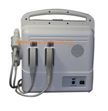 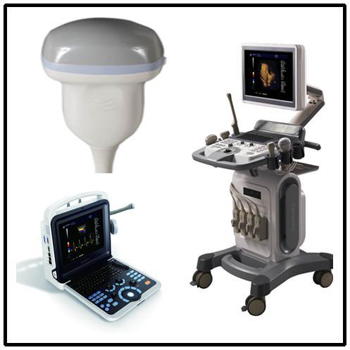
|
 | ||||||||||||
| Â | Â |
WeightGross weight12 kgNet weight7 kg
Â
Power RequirementsVoltage: AC 100V to 240V±10%Frequency: 50Hz±1HzRated Power: 250VAÂ
Operation ConditionsAmbient temperature: 0ºCto +40ºCRelative humidity: 38%  to 85%Atmospheric Pressure: 700hPa to 1060hPaÂ
Software &Â Accessories
Standard AccessoriesPower CableOperation ManualFuseSystem Recovery USB Built in Li-ion batteryÂ
Optional AccessoriesB/W or color Video printerLaserJet or inkjet printerTrolleyAluminum caseBiopsy guideÂ
 Packaging & Delivery
Packaging Details
Gross weight12 kg
Net weight7 kg
YJ-U60PLUS  3D Color Doppler
                --Full Digital Color Doppler Ultrasound Diagnostic System
  Â
Â
AppearanceSmart, compact  and clamshell design12 inch LED monitorBacklit operation panel, 8TGCFloating keyboardTwo active probe connectorsTwo probe holdersÂ
Probe
Transducer Types3.5Mhz convex probe (2.0/ 3.0/ 3.5/ 4.0/ 5.5Mhz)7.5Mhz linear probe (6.0/ 6.5/ 7.5/ 10.0/ 12.0Mhz)3.5Mhz micro convex probe (2.0/ 2.5/ 3.5/4.5/ 5.0Mhz)6.5Mhz transvaginal probe  (5.0/ 6.0/ 6.5/ 7.5/ 9.0Mhz)3.5Mhz phased array probe (2.1/ 3.0/ 3.5/ 4.0/ 5.0Mhz)4D volume probe (2.0/ 3.0/ 3.5/ 4.0/ 5.5Mhz)ApplicationsAbdomen, Obstetrics, Gynecology, Pediatrics,Small parts, Artery,  Superficial organ, Orthopedics, Cardiology, Musculoskeletal, Vascular, etcFunctionAuto Image Optimization Tissue Harmonic imagingiClear (Speckle Noise Reduction)iBeam (Spatial Compound Image)iZoom PIHI (Pulse-Inverse Harmonics Imaging)SA (Synthetic Aperture ultrasonic Imaging)Panoramic Image  (Option)Trapezoid Image (Option)Continuous Wave Doppler(Option)Â
Display modeB, B|B,4B, B|M,M,B|D,PW,B|PW, CFDuplex/Triplex modeCW (option)4D mode (option)Â
ZoomReal time zooming- 10 Steps:  ×1.0, ×2.0, ×3.0, ×4.0, ×5.0, ×6.0, ×7.0, ×8.0, ×9.0, ×10.0Selectable zooming positionÂ
FocusContinuous dynamic focus1~16 selectable transmit focusAcoustic lens focus1, 2, 3, 4 focusÂ
Memory
 Cineâ€memoryBâ€modeMâ€mode SSD (Solid State Disk) 64G
Â
Imaging Processing
B mode8â€step TGC slide pots- Gain: 0~100%
- Depth: 1.6~30cmFrequency: 5Â stepsDynamic range adjustable:Â 0~150dBEdge enhancement:0~7Persistence:0~7Chroma:0~6Grayscale:0~16- Power: 0~100%
- Noise reduction: 0-6iclear: off, 1, 2, 3, 4Â
MÂ mode
- Gain:Â 0~100%Sweep speed: 4Â steps- Maps:Â 0~16Chroma:0~6Â
CÂ mode
- Gain:Â 0~100%Pulse waveWall filter: 4Â stepsColor Maps:Â 0~7Package size: Â 8~15Color persistence:Â 0~7Threshold: 0-3Base line: 0-6Line density: Low and highSpatial filter: 0-3Â
PWÂ mode
- Gain:Â 0~100%Frequency: 5Â stepsPseudo color:0~6PRFd:1.0~6KHzBasic line: 7 stepsWall filter: 7Â steps- Spectrum mode: Refresh and Synchronize
 -   Sampling volume: 0.5-48mm
Â
Measurement &Â Calculation
Measurement
B mode (General) DistanceTrace LengthEllipse (area)Trace(area)AngleVolumeÂ
PW modeHR (heart rate)DistanceVelocityTimeÂ
Calculation
AbdomenLiverGallbladderPancreasSpleen UrologyKidneyUreterBladderAfter the urine bladderProstate GynecologyUterusCervixOvaryFollicle Â
Â
Early Obstetrics GSBPDCRLNT…
Later ObstetricsBPDHCACOFDFLTAD...
Small partsThyroidTestes
Â
MusculoskeletalHipÂ
Peripheral vascularIntimaArteryÂ
CardiologyDistanceAngleVolumeRVWd LVDd RVDd LVPWdRVWsLVDsRVDsLVPWsRV/LVAO…
Â
Physical Features
ConnectivityVideo out portDVI out portVGA out port2  USB portDICOM 3.0Â
Dimension
Â
|
 Full Digital Color Doppler Ultrasound Diagnostic System    AppearanceSmart, compact  and clamshell design12 inch LED monitorBacklit operation panel, 8TGCFloating keyboardTwo active probe connectorsTwo probe holders. Probe Transducer Types3.5Mhz convex probe (2.0/ 3.0/ 3.5/ 4.0/ 5.5Mhz)7.5Mhz linear probe (6.0/ 6.5/ 7.5/ 10.0/ 12.0Mhz)3.5Mhz micro convex probe (2.0/ 2.5/ 3.5/4.5/ 5.0Mhz)6.5Mhz transvaginal probe  (5.0/ 6.0/ 6.5/ 7.5/ 9.0Mhz)3.5Mhz phased array probe (2.1/ 3.0/ 3.5/ 4.0/ 5.0Mhz)4D volume probe (2.0/ 3.0/ 3.5/ 4.0/ 5.5Mhz)ApplicationsAbdomen, Obstetrics, Gynecology, Pediatrics,Small parts, Artery,  Superficial organ, Orthopedics, Cardiology, Musculoskeletal, Vascular, etcFunctionAuto Image Optimization Tissue Harmonic imagingiClear (Speckle Noise Reduction)iBeam (Spatial Compound Image)iZoom PIHI (Pulse-Inverse Harmonics Imaging)SA (Synthetic Aperture ultrasonic Imaging)Panoramic Image  (Option)Trapezoid Image (Option)Continuous Wave Doppler(Option) Display modeB, B|B,4B, B|M,M,B|D,PW,B|PW, CFDuplex/Triplex modeCW (option)4D mode (option) ZoomReal time zooming- 10 Steps:  ×1.0, ×2.0, ×3.0, ×4.0, ×5.0, ×6.0, ×7.0, ×8.0, ×9.0, ×10.0Selectable zooming position FocusContinuous dynamic focus1~16 selectable transmit focusAcoustic lens focus1, 2, 3, 4 focus Memory  Cineâ€memoryBâ€modeMâ€mode SSD (Solid State Disk) 64G  Imaging Processing B mode8â€step TGC slide pots- Gain: 0~100% - Depth: 1.6~30cmFrequency: 5 stepsDynamic range adjustable: 0~150dBEdge enhancement:0~7Persistence:0~7Chroma:0~6Grayscale:0~16- Power: 0~100% - Noise reduction: 0-6iclear: off, 1, 2, 3, 4 M mode - Gain: 0~100%Sweep speed: 4 steps- Maps: 0~16Chroma:0~6 C mode - Gain: 0~100%Pulse waveWall filter: 4 stepsColor Maps: 0~7Package size:  8~15Color persistence: 0~7Threshold: 0-3Base line: 0-6Line density: Low and highSpatial filter: 0-3 PW mode - Gain: 0~100%Frequency: 5 stepsPseudo color:0~6PRFd:1.0~6KHzBasic line: 7 stepsWall filter: 7 steps- Spectrum mode: Refresh and Synchronize  -   Sampling volume: 0.5-48mm  Measurement & Calculation Measurement B mode (General) DistanceTrace LengthEllipse (area)Trace(area)AngleVolume PW modeHR (heart rate)DistanceVelocityTime Calculation AbdomenLiverGallbladderPancreasSpleen UrologyKidneyUreterBladderAfter the urine bladderProstate GynecologyUterusCervixOvaryFollicle   Early Obstetrics GSBPDCRLNT… Later ObstetricsBPDHCACOFDFLTAD... Small partsThyroidTestes  MusculoskeletalHip Peripheral vascularIntimaArtery CardiologyDistanceAngleVolumeRVWd LVDd RVDd LVPWdRVWsLVDsRVDsLVPWsRV/LVAO…  Physical Features ConnectivityVideo out portDVI out portVGA out port2  USB portDICOM 3.0 DimensionGross dimension:510 mm X 500 mm X 330mmNet dimension:330mm X 150 mm X 380mm  Weight Gross weight:12 kg ; Net weight:7 kg  Power RequirementsVoltage: AC 100V to 240V±10%Frequency: 50Hz±1HzRated Power: 250VA Operation ConditionsAmbient temperature: 0ºCto +40ºCRelative humidity: 38%  to 85%Atmospheric Pressure: 700hPa to 1060hPa Software & Accessories Standard AccessoriesPower CableOperation ManualFuseSystem Recovery USB Built in Li-ion battery Optional AccessoriesB/W or color Video printerLaserJet or inkjet printerTrolleyAluminum caseBiopsy guide Probes information Â
Probes informationÂ
Other Products:
|
 | ||||||||||||
| Â | Â |
WeightGross weight12 kgNet weight7 kg
Â
Power RequirementsVoltage: AC 100V to 240V±10%Frequency: 50Hz±1HzRated Power: 250VAÂ
Operation ConditionsAmbient temperature: 0ºCto +40ºCRelative humidity: 38%  to 85%Atmospheric Pressure: 700hPa to 1060hPaÂ
Software &Â Accessories
Standard AccessoriesPower CableOperation ManualFuseSystem Recovery USB Built in Li-ion batteryÂ
Optional AccessoriesB/W or color Video printerLaserJet or inkjet printerTrolleyAluminum caseBiopsy guideÂ
Â
According to the number of barrels, Piston-typed Mobile Milking Machine can be divided into two categories: Single-barrel Type and Double barrel Type.
With unique design, easy operation, easy maintenance, it can lower the utility costs. It will not hurt the nipple by milking soft with stable vacuum. The milking cup group is with a transparent tube, so the operator can observe the nipples of cows during the milking.
After years of production and constant technological improvements, the new designed milking machines can meet the actual demand for the majority of users. We discuss and improve more about the machine, to make them catch international standards, and suitable for more users in different countries. The portable Milking Machine is not only used for the individual animal in small farm, but also provides a selection for large-scale dairy farms for some special animals.
Piston-Typed Milking Machine,Dairy Farm Milking Machine,Piston Milking Machine,Goat Milking Machine
Zibo Maoyang Industry and Trading Co.,ltd , http://www.mysilage.com